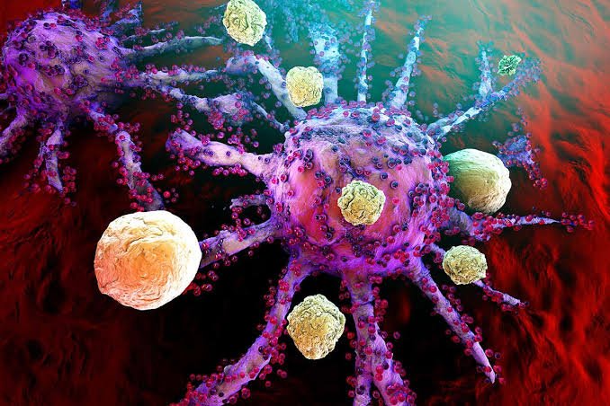
New findings published in Science by researchers from Massachusetts General Hospital (MGH) suggest that immune cells called macrophages may play a crucial role in the development of atrial fibrillation (AFib), a common heart condition characterized by fast and irregular heartbeats. The study analyzed single cells from the atrial heart tissue of AFib patients and found that macrophages were the most dynamic cell population in the atria during AFib, expanding more than any other cell type in diseased tissue.
The researchers created a new mouse model of AFib, referred to as “HOMER,” to investigate the role of macrophages in AFib. They discovered that recruited macrophages contribute to inflammation and scarring (fibrosis) of the atria, impairing the electrical conduction between heart cells and leading to AFib. Inhibiting macrophage recruitment reduced AFib in the mice.
Gene expression analyses revealed that the SPP1 gene was highly overexpressed in macrophages during AFib in both human and mouse hearts. This gene produces the SPP1 protein, also known as osteopontin, which promotes tissue scarring and is elevated in the blood of AFib patients. The researchers observed that HOMER mice lacking this protein had fewer atrial macrophages.
Based on these findings, future therapeutic approaches for AFib could potentially target macrophages or macrophage-derived signals such as SPP1 to counter inflammation and fibrosis. The researchers are actively working on several strategies to develop immunomodulatory therapy for AFib based on these discoveries.
It is crucial to understand how these new strategies could complement existing treatments for AFib. Michelle Olive, Ph.D., from the National Heart, Lung, and Blood Institute, emphasizes the significance of mapping the cardiac and immune cells involved in AFib, as it paves the way for studying how macrophage-targeted therapies can enhance current treatment options.
Source: Massachusetts General Hospital
