Scientists from the University of Tokyo, in collaboration with other researchers, have developed a groundbreaking imaging method called RESORT that allows for unprecedented observation of living systems. Published in the journal Science Advances, this innovative approach combines features from two leading imaging techniques to analyze biological samples in greater detail.
Throughout history, humans have utilized optical devices to examine the microscopic world, constantly striving to improve our understanding by enhancing the tools we use to explore our surroundings, both externally and internally.
Modern microscopic imaging techniques far surpass the capabilities of traditional microscopes. Two prominent technologies in this field are super-resolution fluorescence imaging, which provides high spatial resolution, and vibrational imaging, which sacrifices spatial resolution but allows for labeling multiple constituents in cells using a wide range of colors.
The motivation behind developing RESORT stemmed from the limitations of these existing imaging techniques, prompting researchers to create a superior alternative. Professor Yasuyuki Ozeki from the University of Tokyo’s Research Center for Advanced Science and Technology expressed confidence in their achievement with RESORT: “RESORT stands for reversible saturable optical Raman transitions, and it combines the advantages of super-resolution fluorescence and vibrational imaging without inheriting the drawbacks of either. This laser-based technique utilizes Raman scattering, a special interaction between light and molecules, to identify the components of a sample under the microscope. We have successfully validated the RESORT technique by imaging mitochondria in cells.”
In essence, RESORT represents a significant leap forward in imaging and analysis capabilities, enabling scientists to observe and comprehend living systems with unprecedented clarity and detail.

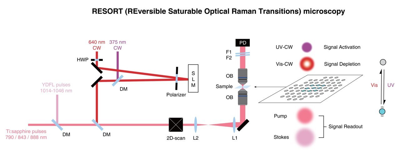
RESORT imaging involves several stages, but the setup is less complex compared to the techniques it aims to replace. The initial step is to label or stain the specific components of the sample with photoswitchable Raman probes, special chemicals that exhibit controlled Raman scattering in response to different laser lights used in RESORT.
Once the sample is labeled, it is placed within an optical apparatus that illuminates it appropriately and constructs an image. This involves irradiating the sample with two-color infrared laser pulses to detect Raman scattering, as well as utilizing ultraviolet light and a donut-shaped beam of visible light. Together, these elements confine the area where Raman scattering occurs, enabling high spatial resolution during the imaging stage.
According to Professor Ozeki, the significance of RESORT goes beyond achieving higher-resolution images of microscopic samples, as electron microscopes can already provide greater detail. The advantage lies in the fact that electron microscopes inherently damage or hinder the samples they observe. By expanding the range of colors available for Raman probes, RESORT has the potential to image multiple components within living samples, capturing dynamic interactions and facilitating a deeper understanding of fundamental biological processes, disease mechanisms, and potential therapeutic interventions.
While the primary focus of the team was to enhance microscopic imaging for medical research and related fields, the advancements in laser design achieved through RESORT could find applications in other areas that require high power or precise control, such as materials science.
Source: University of Tokyo